The cells are mixed with extracellular matrix material derived from the patient’s own tissue, forming a natural hydrogel that mimics the environment cells experience inside the body. This bio-ink provides biochemical cues that guide cell behavior, growth, and organization
What Israeli scientists at Tel Aviv University have achieved with the 3D printing of a living, human-cell-based heart marks one of those rare moments in science where the future seems to briefly step into the present. It is not yet a future where surgeons routinely replace failing hearts with freshly printed ones, nor is it a miracle cure that makes donor shortages instantly disappear. But it is a moment where decades of theoretical promise in regenerative medicine, stem cell biology, and bioengineering have converged into a tangible, beating object—small, imperfect, and experimental, yet profoundly symbolic.
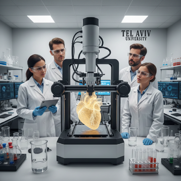
The research team, led by Professor Tal Dvir at Tel Aviv University, has demonstrated for the first time that it is possible to fabricate an entire heart structure using only human biological material derived from a patient’s own body. Unlike earlier tissue engineering efforts that focused on patches, valves, or simplified tissue scaffolds, this work attempts something far more ambitious: printing a whole organ complete with chambers, blood vessels, and cardiac muscle capable of contracting. Even though the resulting heart is only about the size of a rabbit’s and cannot yet pump blood effectively, its existence fundamentally alters what scientists believe is possible.
The process begins with something deceptively simple—a small biopsy of fat tissue taken from the patient. Fat, long treated as an undesirable byproduct of modern lifestyles, becomes the raw material for one of the most sophisticated medical technologies ever attempted. From this biopsy, scientists extract cells and reprogram them into pluripotent stem cells, which have the remarkable ability to become almost any cell type in the human body. These stem cells are then coaxed into differentiating into cardiac muscle cells, endothelial cells for blood vessels, and other supporting cell types necessary for heart tissue.
What makes this approach especially significant is the creation of what researchers call “bio-ink.” The cells are mixed with extracellular matrix material derived from the patient’s own tissue, forming a natural hydrogel that mimics the environment cells experience inside the body. This matrix is not merely a passive scaffold; it provides biochemical cues that guide cell behavior, growth, and organization. When loaded into a specialized 3D bioprinter, this bio-ink can be deposited layer by layer with extraordinary precision, allowing scientists to recreate the complex architecture of a heart rather than just a simple lump of tissue.
The printing itself is an engineering feat, but the true challenge lies in coaxing the printed structure into behaving like living heart tissue. The heart is not just a mass of muscle; it is an exquisitely coordinated machine in which electrical signals, mechanical contraction, and blood flow must work in harmony. The Tel Aviv University team has shown that their printed heart tissue can contract spontaneously, a crucial milestone that distinguishes living cardiac tissue from inert biological constructs. However, coordinated pumping—the ability to move blood efficiently through chambers and vessels—remains an unsolved problem at this stage.
It is important to emphasize what this achievement is and what it is not. Online narratives and social media posts have sometimes exaggerated the breakthrough, suggesting that a fully functional human heart has already been created and is ready for transplantation. That is not the case. This heart is a proof of concept, a demonstration that the fundamental biological and engineering principles required for personalized organ printing can work together. It is closer to a prototype than a finished product. Yet in medical research, proof of concept is often the most difficult step, because it establishes that a problem once thought nearly impossible is, in fact, solvable.
The implications for transplantation medicine are immense. Today, heart transplantation remains the only definitive treatment for end-stage heart failure, a condition that affects millions worldwide. Donor organs are desperately scarce, and waiting lists are long. In the United States alone, an average of 18 people die each day while waiting for a suitable organ. Globally, the numbers are even more sobering. Even when a donor heart becomes available, patients must undergo lifelong immunosuppressive therapy to prevent rejection, exposing them to infections, cancer, and other serious complications.
A heart grown from a patient’s own cells offers a radically different paradigm. Because the tissue is genetically identical to the recipient, the risk of immune rejection could be dramatically reduced or even eliminated. This would not only improve survival rates but also enhance quality of life by reducing dependence on powerful immunosuppressive drugs. In theory, it could transform transplantation from a desperate race against time into a planned, personalized medical procedure.
Beyond transplantation, the technology opens doors in drug development and disease research. Lab-grown human heart tissue can serve as a testing ground for new medications, allowing researchers to observe how real human cardiac cells respond to drugs without risking patient safety. This could significantly reduce the reliance on animal testing, which often fails to accurately predict human responses. Personalized heart tissue could also be used to study genetic heart diseases, enabling doctors to test treatments on a patient’s own cells before prescribing them.
The road from a rabbit-sized, non-pumping heart to a full-scale, transplantable human organ, however, is long and filled with challenges. Scaling up is not simply a matter of making the printer larger. As organs grow in size, ensuring adequate oxygen and nutrient delivery to every cell becomes increasingly difficult. In the human body, this problem is solved by an intricate network of blood vessels ranging from large arteries to microscopic capillaries. Replicating this vascular complexity in a printed organ is one of the greatest hurdles in regenerative medicine.
Another challenge lies in maturation. The cells in printed heart tissue often resemble those of a developing or fetal heart rather than an adult one. They can contract, but not with the strength, endurance, or coordination required for sustained pumping over years or decades. Researchers are exploring various strategies to promote maturation, including electrical stimulation, mechanical conditioning, and biochemical cues that mimic the environment of a beating heart inside the body.
Ethical and regulatory questions also loom large. The idea of printing human organs raises issues about access, cost, and equity. Will such technologies be available only in elite hospitals in wealthy countries, or can they be scaled and standardized to benefit patients globally? How will regulatory agencies evaluate the safety and effectiveness of organs that are, by definition, unique to each patient? These are not merely technical questions but societal ones that will shape how regenerative medicine is integrated into healthcare systems.
Professor Tal Dvir himself has been careful to balance optimism with realism. His vision of organ printers becoming standard equipment in leading hospitals within a decade captures the excitement of the field while acknowledging the work still to be done. The timeline may shift as unexpected challenges arise, but the direction is clear. What once belonged to the realm of science fiction is steadily becoming a branch of clinical science.
Heart disease remains the leading cause of death in many parts of the world, driven by aging populations, sedentary lifestyles, and metabolic disorders. Traditional approaches—medications, lifestyle interventions, mechanical assist devices, and transplantation—have saved countless lives, but they also have inherent limits. The possibility of replacing damaged organs with living, personalized replacements represents a fundamental shift from managing disease to restoring function.
In this context, the Tel Aviv University heart is more than a scientific curiosity. It is a statement about the future of medicine, one in which biology is not merely repaired but re-engineered with precision. It challenges the long-standing assumption that organ failure is irreversible and that scarcity must always dictate who receives life-saving treatment.
At the same time, it serves as a reminder of the importance of scientific rigor and honest communication. Breakthroughs of this magnitude attract hype, but progress in medicine is incremental, built on careful experimentation and repeated validation. The small, silent heart printed in a laboratory today does not yet beat for a patient, but it beats loudly as an idea—an idea that the body’s own cells, guided by human ingenuity, can be persuaded to rebuild what disease has destroyed.
If the next decade fulfills even part of the promise suggested by this research, hospitals may one day house not only operating theaters and intensive care units but also biofabrication labs where organs are grown to order. Such a transformation would redefine transplantation, reshape medical economics, and alter our understanding of what it means to heal. The printed heart from Tel Aviv University stands at the threshold of that transformation, imperfect but extraordinary, a quiet herald of a future in which the line between biology and technology grows ever thinner.
The creation of a living, three-dimensional, human heart using a patient’s own biological material represents one of the most consequential advances in regenerative medicine in recent decades. At Tel Aviv University, a team of Israeli scientists led by Professor Tal Dvir has demonstrated, for the first time, the feasibility of printing an entire heart-like structure composed of human cells, complete with chambers, blood vessels, and cardiac tissue capable of contraction. Although this organ is currently small—roughly the size of a rabbit’s heart—and lacks the ability to pump blood in a fully functional manner, the achievement marks a critical proof of concept. It signals a future in which personalized, lab-grown organs may fundamentally reshape the treatment of heart disease and, more broadly, the global transplant ecosystem.
Heart disease remains the leading cause of death worldwide, placing an immense burden on healthcare systems and families alike. For patients with end-stage heart failure, transplantation is often the only viable option. Yet the supply of donor hearts falls dramatically short of demand. In the United States alone, thousands of patients wait anxiously on transplant lists, and on average, around eighteen people die each day before a suitable organ becomes available. Similar shortages exist across Europe, Asia, and much of the developing world. Against this backdrop, the idea of manufacturing a patient-specific heart on demand has long occupied the realm of theoretical science and speculative medicine. The work emerging from Tel Aviv University brings that idea closer to tangible reality.
At the heart of this breakthrough is the innovative use of a patient’s own fat tissue as the foundational biological material. The process begins with a simple biopsy, during which a small sample of adipose tissue is extracted from the patient. Fat cells, once regarded as biologically mundane, have in recent years gained prominence as a rich and accessible source of cellular material. From this biopsy, scientists isolate living cells and reprogram them into pluripotent stem cells—cells with the capacity to differentiate into virtually any tissue type in the human body. This reprogramming step is critical, as it allows the same original cells to give rise to heart muscle cells, blood vessel cells, and other supporting tissues necessary for organ construction.
Once reprogrammed, these cells are combined with an extracellular matrix derived from the patient’s own tissue. This matrix functions as a natural scaffold, forming a hydrogel that supports cell growth and organization. The resulting substance, often referred to as “bio-ink,” is biologically compatible and uniquely personalized. Unlike synthetic materials or donor-derived tissues, it carries the patient’s own molecular signature, thereby eliminating the immune incompatibilities that typically plague organ transplantation. In conventional transplants, the recipient’s immune system frequently recognizes the new organ as foreign, necessitating lifelong immunosuppressive therapy with significant side effects. By contrast, organs printed from a patient’s own cells theoretically remove the risk of rejection altogether.
The bio-ink is then fed into a specialized 3D bioprinter designed to deposit living material with extraordinary precision. Layer by layer, the printer constructs the architecture of the heart, including chambers, valves, and a network of blood vessels. This vascular component is particularly significant, as it represents one of the greatest challenges in tissue engineering. Many earlier attempts at growing organs failed because the tissues could not be adequately supplied with oxygen and nutrients. By incorporating blood vessels directly into the printed structure, the Tel Aviv University team addressed a long-standing bottleneck in the field.
The result is a miniature heart-like organ that closely resembles its natural counterpart in structure and composition. Importantly, the cardiac tissue within this printed heart is capable of spontaneous contraction, meaning it can beat independently under laboratory conditions. While this beating is not yet coordinated or forceful enough to pump blood effectively, it demonstrates that the cells are not merely alive but functionally active. This distinction elevates the achievement from simple tissue fabrication to genuine organ-level engineering.
Despite the significance of this development, Professor Dvir and his colleagues have been careful to temper public expectations. In an era of rapid information sharing and social media amplification, scientific breakthroughs are often exaggerated or misrepresented. Claims that a fully functional human heart has already been printed and is ready for transplantation are inaccurate. The current organ is too small, too weak, and insufficiently mature to sustain human life. Rather than a finished product, it is best understood as a sophisticated prototype—a demonstration that the fundamental principles required for personalized organ printing are sound.
The challenges that remain are formidable. Scaling the heart from rabbit size to full human dimensions is not simply a matter of increasing volume. Larger organs require more complex vascular networks, stronger and more synchronized muscle contractions, and advanced electrical signaling systems to coordinate heartbeat rhythm. Additionally, printed tissues must undergo a maturation process that mimics natural development, allowing cells to align properly and acquire the mechanical strength necessary for continuous pumping over many years. Researchers are actively exploring ways to stimulate this maturation through mechanical conditioning, electrical stimulation, and biochemical cues.
Beyond transplantation, the implications of this technology extend into other critical areas of medicine. Lab-grown heart tissues offer unprecedented opportunities for drug testing and disease modeling. Pharmaceutical development is notoriously expensive and risky, in part because drugs that appear safe in animal models can have unexpected effects in humans. By testing new compounds on human cardiac tissue derived from real patients, researchers can gain earlier and more accurate insights into drug safety and efficacy. This approach could reduce reliance on animal testing and accelerate the development of treatments for heart disease and other conditions.
Personalized organ printing also opens new avenues for studying genetic and acquired heart disorders. Tissues created from patients with specific diseases can be observed under controlled conditions, allowing scientists to investigate disease progression at a cellular level. Such insights could lead to more targeted therapies and earlier interventions, potentially preventing heart failure before it reaches an advanced stage.
From an ethical and societal perspective, the prospect of printed organs raises important questions. If organ printers become a standard feature in major hospitals, as Professor Dvir has suggested might be possible within a decade, issues of access and equity will come sharply into focus. Will these technologies be available only in wealthy nations and elite medical centers, or can they be scaled affordably to serve broader populations? How will healthcare systems regulate and prioritize their use? While these questions remain unresolved, the technology itself offers hope for a more humane and efficient alternative to the current transplant system, which relies on tragedy and scarcity.
The Israeli team’s achievement also highlights the growing role of interdisciplinary collaboration in modern science. This work sits at the intersection of molecular biology, materials science, biomedical engineering, and clinical medicine. Advances in one domain—such as stem cell reprogramming or 3D printing precision—enable breakthroughs in another. Tel Aviv University’s success underscores the importance of sustained investment in basic research, as many of the underlying techniques were developed years or even decades before their full potential became apparent.
In the broader global context, regenerative medicine is rapidly evolving, with research groups across the United States, Europe, and Asia pursuing parallel approaches to organ engineering. What distinguishes the Tel Aviv University heart is its completeness: it is not merely a patch of cardiac tissue or a partial structure but an entire organ framework printed from human cells. This holistic approach sets a new benchmark for the field and provides a clear roadmap for future efforts.
Ultimately, the significance of this breakthrough lies not in what it delivers today but in what it makes possible tomorrow. It represents a shift from the idea of organs as scarce, donated resources to organs as customizable, patient-specific constructs. While many technical and regulatory hurdles remain, the fundamental question—whether a human heart can be printed from a patient’s own cells—has been answered in the affirmative, at least in principle.
As Professor Tal Dvir has noted, it is conceivable that within ten years, organ printers could become a routine presence in leading hospitals around the world. If that vision is realized, the impact on human health would be profound. Countless lives currently lost to heart failure and transplant shortages might be saved, and the very definition of medical treatment would expand to include the on-demand creation of living replacement organs. The small, beating heart produced in a Tel Aviv laboratory may thus be remembered not for its size or immediate utility, but as the first tangible step toward a future in which regenerative medicine fulfills its most ambitious promise.